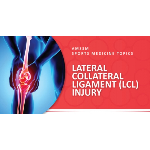
 What is it? A ligament is a short band of tough fibrous connective tissue that holds two bones together. In the knee, there are four major ligaments: anterior cruciate ligament (ACL), posterior cruciate ligament (PCL), medial collateral ligament (MCL), and the lateral collateral ligament (LCL). The LCL is located on the outer part of the knee and connects the femur (thigh bone) and fibula (smaller of the two leg bones). It can get injured or torn when the knee twists or more commonly when hit from the inside out (also known as varus stress mechanism). Compared to the MCL, the LCL is less commonly injured because the anatomy is complex on the outside of the knee. It accounts for only 2% of knee injuries in isolation. Most LCL injuries also include injuries to the posterior lateral corner, posterior cruciate ligament (PCL), lateral meniscus, or anterior cruciate ligament (ACL). Symptoms/Risks • Symptoms of lateral collateral ligament injury may include: Pain and swelling on the outside or posterolateral aspect of the knee • Pain with walking or bending the knee • Feelings of instability (knee giving out) while walking or doing activity • Mechanical symptoms (locking, catching) may indicate associated meniscus injury Risks associated with LCL injury: - No specific risks for LCL injury have been identified - Risks for general knee injury include: - Female gender - Competitive play (not practice) - Injury due to player-to-player contact - Sports that involve pivoting, jumping, landing
Sports Medicine Evaluation & Treatment A sports medicine physician will take your complete history on how the injury occurred and perform a physical exam on the knee. In the physical exam, he or she will inspect the knee looking for obvious deformities, range of motion, swelling, bruises or collection of blood. The physician will then examine the knee. The most common exam finding in patients with an LCL injury is tenderness along the lateral knee at the site where the patient often complains of pain. To assess your LCL injury, he or she will perform a varus stress test – this involves the physician pushing on the inside of the knee with the knee fully extended and with the knee bent at a 30 degree angle to assess for any laxity of the LCL. The physician may order an x-ray to make sure there is no bony damage from the injury. An MRI is not usually needed for these types of injuries unless there is a suspicion for other ligament or meniscus injuries. However, MRI is the diagnostic test of choice to assess the extent of damage of the LCL Most isolated LCL injuries without other ligament or meniscus injuries heal well without surgery. Typically, you can do the following: • Apply ice for 20 minutes three times a day to help with pain and swelling. • Take anti-inflammatory medications such as Ibuprofen to help with pain. • Try a hinged knee brace to help stabilize the knee while it heals. • A referral to physical therapy to help strengthen the knee will be made by your physician • A sports medicine physician may also inject the knee to help decrease inflammation to accelerate healing • If there are other injuries along with the LCL injury or the knee is unstable, surgery may be considered.
Injury Prevention Most LCL injuries happen because of trauma or accident. A good knee-strengthening and core muscle program may decrease the risk for injury, but these programs not completely prevent it. The physician will teach you some of these exercises and give you information on how to do them at home.
Return to Play Most athletes with an LCL injury not requiring surgery return to sports within 6-8 weeks. If surgery is needed, the type of surgery, the number of ligaments injured/ repaired, as well as the rehabilitation time required will determine length of time before return to play. The sports medicine physician works in conjunction with coaches and athletic trainers to make this transition possible. AMSSM Member Authors References Category: Leg and Thigh, Overuse Injuries, [Back] |

