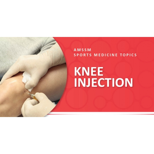
 What is it? The knee joint divided up into three compartments that share joint fluid and come together to make one larger joint cavity. These compartments are the patellofemoral, lateral tibiofemoral, and medial tibiofemoral compartments. Patients can develop different problems in the knee joints that may require injections for diagnostic and therapeutic purposes. One of the most common problem is knee osteoarthritis (OA), which is a degenerative process from wear and tear over time on the joint. Treatment guidelines for knee OA recommend a combination of non-pharmacological and pharmacological treatments, which may include procedures in the joint. Intraarticular injections can help reduce pain and inflammation of the knee joint. These can help lead to better movement, performance of daily activities, exercising, and/ or rehabilitation. Most commonly, a solution of an anesthetic and a corticosteroid is used. Alternative injection options include viscosupplementation and regenerative products. There has been an increasing trend in using regenerative medicine therapies to treat knee OA. These products include prolotherapy, platelet rich plasma (PRP), bone marrow aspirate concentrate (BMAC), adipose-derived stem cells (ASCs), and amniotic tissue products. The research evidence behind these products is variable; as such, we recommend discussing these options with physicians prior to considering them. Procedure Details The knee joint cavity can be injected from a variety of different places around the knee. X-rays can be helpful to determine which compartment has the least obstruction in order to get the needle into the joint. Ultrasound guidance can be a good and accurate tool to assist with injections in patients where traditional injection techniques might be challenging. The most common approaches for knee joint injections are superolateral (above and outside of the kneecap, also known as the patella), superomedial (above and to the middle of the patella), anteromedial (below and to the middle of the patella), and anterolateral (above and outside of the patella). The superolateral approach may be a more reliable route of entry to reach the inside of the capsule (joint). With the superolateral and superomedial approaches, the patient lies on their back with the knee extended. Starting at the center of the patella, the needle is inserted and directed slightly toward the back of the knee joint with the intention of the needle behind directly behind the patella. On the other hand, the anterolateral and anteromedial approaches are done with the patient seated, with the knee hanging off the edge of the table. The needle is inserted just below the patella on the medial or lateral side of the patellar tendon and directed toward the center of the knee joint. Before the needle is inserted, the selected injection spot is marked and cleaned before the procedure. Every precaution is taken to keep the area clean/sterile. Cold spray may be used on occasion to numb the skin before injecting. The needle will be introduced until it reaches the joint space as described above. The injection should flow easily into the knee joint. The risk of bleeding is minimal; however, special attention may be taken with patient taking blood thinners. Ultrasound guidance can be used to improve accuracy and minimize pain. Ultrasound may also be indicated: 1) after failure of a prior traditional injection technique as described above; 2) a patient with obesity that makes traditional techniques challenging 3) a need for diagnostic specificity, and; 4) orthobiologic injections in which accuracy is essential for the treatment. Post-Procedure Guidance The knee may feel better within minutes if anesthetic medication was used in the injection, with the effect lasting a few hours. The effect of the steroid medication may be seen between 2-7 days after the procedure. Rarely, the pain can increase on the first 24-48 hours due to a phenomenon called “steroid flare.” This will improve on its own but can be managed with ice, nonsteroidal anti-inflammatory drugs (NSAIDS), or acetaminophen. After the procedure, the knee must not be soaked or submerged in water for 48 hours. Possible complications from this procedure include infection, skin discoloration, and tendon injury. Special consideration should be given to diabetic patients because steroid injections can elevate blood sugars for a few days. Diabetic patients should monitor their glucose levels frequently for the first few days. In general, complications from knee intra-articular injections are rare, especially if the proper technique and necessary sterile precautions were taken. Return to Play After the injection, patient should avoid rigorous activity for 24-48 hours. There is a small but potential risk of tendon injury after an injection. That is why, sometimes, steroid injections are avoided for athletes who are in season for their sport or have upcoming competitions. AMSSM Member Authors References Category: Knee, Treatments in Sports Medicine, [Back] |

